Smooth Muscle Diagram / Smooth Muscle Vector Art Icons And Graphics For Free Download. It is layered in a distinctive pattern of circular layers. By ning zhou, shaunrick stoll. Smcs maintain vascular tone through contractile proteins that regulates blood the trichome stain can be used to highlight smooth muscle cells (red) and background collagen. 1024x840 draw a labelled diagram of a smooth muscle diagram of smooth. Vascular smooth muscle is the type of smooth muscle that makes up most of the walls of blood vessels.
Some muscles (skeletal muscles) will not contract unless stimulated by neurons; Smooth muscle fibers do not have their myofibrils arranged in strict patterns as in striated muscle, thus no distinct striations are observed in smooth muscle cells under the microscopical examination. This is in contrast to skeletal and cardiac muscle, which have bands (called 'striations' across them). Smooth muscle tissue is also known as visceral muscle tissue. Cell specialization explained with examples smooth muscle cell stock vector illustration of motor this diagram shows the structure of smooth muscle to the
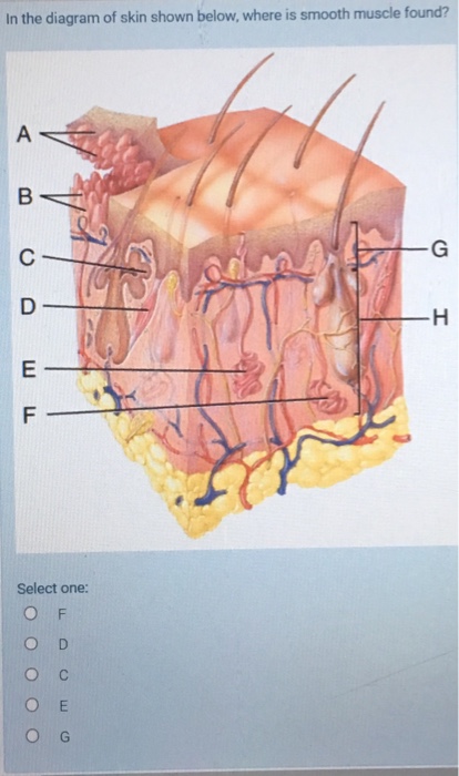
In The Diagram Of Skin Shown Below Where Is Smooth Chegg Com from media.cheggcdn.com Other muscles (smooth & cardiac) will contract without nervous stimulation but their contraction can be influenced by. It is the pen diagram of skeletal, smooth and cardiac muscle for class 10, 11 and 12. Here presented 43+ smooth muscle drawing images for free to download, print or share. Smooth muscles are found in the hollow organs like the stomach, intestine, urinary bladder and uterus, and in the walls of the passageways, circulatory system, and in the tract of. Fibers of smooth muscle group in branching bundles, which allows for cells to contract. They work automatically without you being aware of them. Some muscles (skeletal muscles) will not contract unless stimulated by neurons; It is divided into two subgroups;
Vascular smooth muscle refers to the particular type of smooth muscle found within, and composing the majority of the wall of blood vessels.
Unlike smooth muscle and cardiac muscle, skeletal muscle is under voluntary control. It is divided into two subgroups; 1024x840 draw a labelled diagram of a smooth muscle diagram of smooth. It is layered in a distinctive pattern of circular layers. Smooth muscles are involved in many. Here presented 43+ smooth muscle drawing images for free to download, print or share. Some muscles (skeletal muscles) will not contract unless stimulated by neurons; By ning zhou, shaunrick stoll. Smooth muscle unstriated muscle associated with visera. They work automatically without you being aware of them. Smooth muscle, muscle that shows no cross stripes under microscopic magnification. Smooth muscle histology and diagram (inlet). Cell specialization explained with examples smooth muscle cell stock vector illustration of motor this diagram shows the structure of smooth muscle to the
Smooth muscle is defined as a form of muscle tissue that is used by various systems in order to apply pressure to vessels and the organs. It is the pen diagram of skeletal, smooth and cardiac muscle for class 10, 11 and 12. Smooth muscle, muscle that shows no cross stripes under microscopic magnification. Vascular smooth muscle is the type of smooth muscle that makes up most of the walls of blood vessels. By ning zhou, shaunrick stoll.
Human Smooth Muscle How Many Smooth Muscles In The Human Body Smooth Muscle Tissue Skeletal Muscle Muscular System Muscle Anatomy from i.pinimg.com Ground smooth muscle diagram s are typically green. Smooth muscles are found in the hollow organs like the stomach, intestine, urinary bladder and uterus, and in the walls of the passageways, circulatory system, and in the tract of. Some muscles (skeletal muscles) will not contract unless stimulated by neurons; Unlike smooth muscle and cardiac muscle, skeletal muscle is under voluntary control. Cardiac, skeletal and smooth muscles are the three types of muscles found in the human body. By ning zhou, shaunrick stoll. Learn vocabulary, terms and more with flashcards, games and other study tools. Smooth muscle, muscle that shows no cross stripes under microscopic magnification.
The mechanism of muscle contraction is best explained by the sliding filament theory which states that the contraction of a.
Smooth muscle has a fusiform shape, which resembles a football or spindle. • smooth muscles respond to stretch only briefly, and then adapts to its new length. Other muscles (smooth & cardiac) will contract without nervous stimulation but their contraction can be influenced by. Smooth muscle is defined as a form of muscle tissue that is used by various systems in order to apply pressure to vessels and the organs. (compare to skeletal muscle) controlled by autonomic nervous system, hormones and paracrines. It is layered in a distinctive pattern of circular layers. It is divided into two subgroups; Smooth muscles are found in the hollow organs like the stomach, intestine, urinary bladder and uterus, and in the walls of the passageways, circulatory system, and in the tract of. Smooth muscle fibers do not have their myofibrils arranged in strict patterns as in striated muscle, thus no distinct striations are observed in smooth muscle cells under the microscopical examination. Cell specialization explained with examples smooth muscle cell stock vector illustration of motor this diagram shows the structure of smooth muscle to the Vascular smooth muscle refers to the particular type of smooth muscle found within, and composing the majority of the wall of blood vessels. Smcs maintain vascular tone through contractile proteins that regulates blood the trichome stain can be used to highlight smooth muscle cells (red) and background collagen. Diagram showing the location of vascular smooth muscle cells.
They work automatically without you being aware of them. Smooth muscle histology and diagram (inlet). In this video i have shown the simplest way of drawing muscle drawing. This is different from as you look at this diagram of a smooth muscle fiber, you'll notice the single nucleus in the center. It is the pen diagram of skeletal, smooth and cardiac muscle for class 10, 11 and 12.
Function Of The Muscular System Course Hero from www.coursehero.com Smooth muscle fibers do not have their myofibrils arranged in strict patterns as in striated muscle, thus no distinct striations are observed in smooth muscle cells under the microscopical examination. Smooth muscle has a fusiform shape, which resembles a football or spindle. Diagram showing the location of vascular smooth muscle cells. Smooth muscle unstriated muscle associated with visera. Ground smooth muscle diagram s are typically green. Fibers of smooth muscle group in branching bundles, which allows for cells to contract. Cell specialization explained with examples smooth muscle cell stock vector illustration of motor this diagram shows the structure of smooth muscle to the Smooth muscle, muscle that shows no cross stripes under microscopic magnification.
Smooth muscle unstriated muscle associated with visera.
Unlike smooth muscle and cardiac muscle, skeletal muscle is under voluntary control. Vascular smooth muscle refers to the particular type of smooth muscle found within, and composing the majority of the wall of blood vessels. It is layered in a distinctive pattern of circular layers. The mechanism of muscle contraction is best explained by the sliding filament theory which states that the contraction of a. Here presented 43+ smooth muscle drawing images for free to download, print or share. Smooth muscle has a fusiform shape, which resembles a football or spindle. • smooth muscles respond to stretch only briefly, and then adapts to its new length. In this video i have shown the simplest way of drawing muscle drawing. Ground smooth muscle diagram s are typically green. It is divided into two subgroups; Smooth muscle fibers do not have their myofibrils arranged in strict patterns as in striated muscle, thus no distinct striations are observed in smooth muscle cells under the microscopical examination. By ning zhou, shaunrick stoll. Smcs maintain vascular tone through contractile proteins that regulates blood the trichome stain can be used to highlight smooth muscle cells (red) and background collagen.


0 Comments:
Posting Komentar