Home
Uncategories
Pelvic Anatomy Ligaments - 32. The anatomy, histology and development of the uterine tube and uterus. | DOTE Anatomy topics / Anatomynote.com found pelvis and ligaments front view from above male from plenty of anatomical pictures on the internet.
Pelvic Anatomy Ligaments - 32. The anatomy, histology and development of the uterine tube and uterus. | DOTE Anatomy topics / Anatomynote.com found pelvis and ligaments front view from above male from plenty of anatomical pictures on the internet.
Pelvic Anatomy Ligaments - 32. The anatomy, histology and development of the uterine tube and uterus. | DOTE Anatomy topics / Anatomynote.com found pelvis and ligaments front view from above male from plenty of anatomical pictures on the internet.. Anatomynote.com found pelvis and ligaments front view from above male from plenty of anatomical pictures on the internet. This image added by admin. The sacral ligaments are responsible for the major connection between the three bones of the pelvis. ^^^these two ligaments are from ovarian gubernaculum; This will be explored further on.
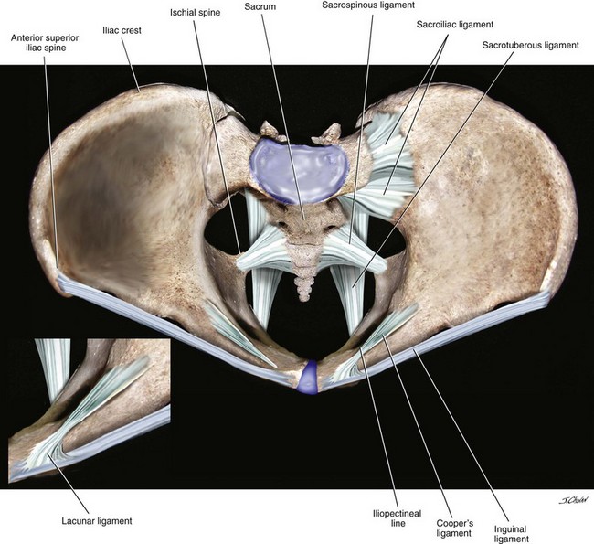
These ligaments firmly hold together the two pubic bones and, consequently, the two innominate bones. The pelvic girdle and pelvic spine. Cookies allow us to analyze and store information such as the characteristics of your device as well as certain personal data (e.g., ip addresses, navigation, usage or geolocation data, unique identifiers). There are ligaments between the sacrum and the ilium, which are called sacroiliac ligaments. The outlet is formed by the pubic arch, ischial spines, sacrotuberous ligaments, and the coccyx.
The Inguinal Canal - Boundaries - Contents - TeachMeAnatomy from s3.amazonaws.com ^^^these two ligaments are from ovarian gubernaculum; The broad ligament is a sheet of pelvic peritoneum extending bilaterally from the lateral pelvic sidewalls to the uterus in the midline. The enclosed space between the inlet and outlet is called the true pelvis, with the plane of the inlet being at right angles to the plane of the outlet. It is close to the major vasculature of the pelvis, including external iliac vein. The inlet to the pelvic canal is at the level of the sacral promontory and superior aspect of the pubic bones. The pectineal ligament is strong, and holds suture well. The broad ligament folds over the fallopian tubes and ovaries and covers them anteriorly and posteriorly. There are 2 round ligaments, one on each side of the uterus.
The ligaments that connect the sacrum to the coccyx.
The outlet is formed by the pubic arch, ischial spines, sacrotuberous ligaments, and the coccyx. Explore every muscle, bone and organ in 3d The three bony structures (ilium, ischium, and pubis) are held together with strong ligaments that are important in understanding pelvic anatomy and biomechanics. There are ligaments in front of the pubic bones where they are next to each other. The pelvic girdle and pelvic spine. During pregnancy, the pelvic joints and ligaments are relaxed, so that the relaxed, so that the range of motion is increased and the locking mechanism becomes less efficient. Iliolumbar, sacrotuberous and sacrospinous ligaments. The sacroiliac strain thus produced may persist even after pregnancy. The broad ligament is a flat sheet of peritoneum, associated with the uterus, fallopian tubes and ovaries. These ligaments firmly hold together the two pubic bones and, consequently, the two innominate bones. It extends from the lateral pelvic walls on both sides, and folds over the internal female genitalia, covering their surface anteriorly and posteriorly. You can click the image to magnify if you cannot see clearly. The ilium, ischium and the pubic bone.
Imaios and selected third parties, use cookies or similar technologies, in particular for audience measurement. Anatomy app 3d unlocks the world of human body. Study human anatomy with reliable 3d models & detailed articles. The most important pelvic ligaments are as follows: Laterally, the broad ligament is prolonged over the vessels as the suspensory ligament of the ovary
Surgical Anatomy of the Pelvis and the Anatomy of Pelvic Support | Abdominal Key from abdominalkey.com The pectineal ligament is usually around 6 cm long in adults. The enclosed space between the inlet and outlet is called the true pelvis, with the plane of the inlet being at right angles to the plane of the outlet. The broad ligament folds over the fallopian tubes and ovaries and covers them anteriorly and posteriorly. Those that connect the ilium to the sacrum; These ligaments are categorized into four groups: This naturally puts a greater strain on the ligaments. There are two major groups of ligaments that provide nearly all the structure of the pelvis. All axis rotatable modeled 3d model pelvis and ligaments, labeled.
The ligaments that connect the sacrum to the coccyx.
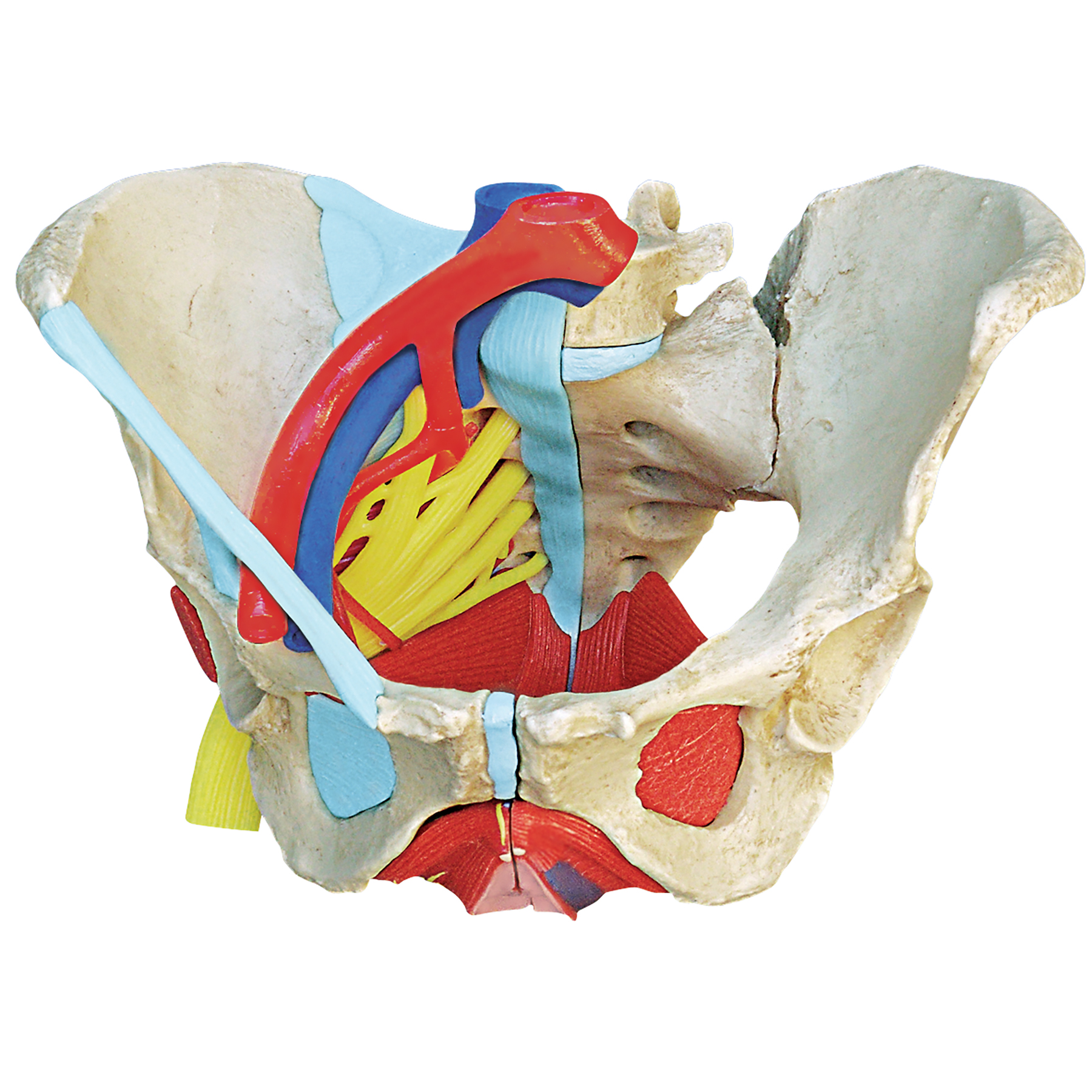
The pelvis consists of two innominate bones and the sacrum to which coccyx is attached. All axis rotatable modeled 3d model pelvis and ligaments, labeled. The sacral ligaments are responsible for the major connection between the three bones of the pelvis. The pelvic girdle, also known as the hip bone, is composed of three fused bones: Anatomynote.com found pelvis and ligaments front view from above male from plenty of anatomical pictures on the internet. The sacroiliac strain thus produced may persist even after pregnancy. If you would like a large, unwatermarked image for your web page or blog, please purchase the appropriate license. The enclosed space between the inlet and outlet is called the true pelvis, with the plane of the inlet being at right angles to the plane of the outlet. They are traversed by the ureters and pelvic blood vessels. Pelvic floor consists of two ligaments: There are ligaments in front of the pubic bones where they are next to each other. The ligaments that connect the sacrum to the coccyx. The femoral ligaments act to stabilize the ball and socket joint of the hip, connecting to the ilium and the ischium.
Study human anatomy with reliable 3d models & detailed articles. They are traversed by the ureters and pelvic blood vessels. The pelvic ligaments are strong, thick bands of fibrous tissue that connect the pelvic bones. The most important pelvic ligaments are as follows: We hope you can get the exact.
Anatomical Female Pelvis Model | Childbirth Graphics from www.childbirthgraphics.com This will be explored further on. Sacrotuberous ligament sacrospinous ligament lesser sciatic foramen greater sciatic foramen medial attachment: Medial surface of greater trochanter innervation: If you would like a large, unwatermarked image for your web page or blog, please purchase the appropriate license. Thank you for visit anatomynote.com. The broad ligament is a sheet of pelvic peritoneum extending bilaterally from the lateral pelvic sidewalls to the uterus in the midline. The pelvic ligaments are strong, thick bands of fibrous tissue that connect the pelvic bones. The pectineal ligament is strong, and holds suture well.
If you would like a large, unwatermarked image for your web page or blog, please purchase the appropriate license.
The broad ligament is a sheet of pelvic peritoneum extending bilaterally from the lateral pelvic sidewalls to the uterus in the midline. The pelvis is held together by three principal ligaments: The broad ligament is a flat sheet of peritoneum, associated with the uterus, fallopian tubes and ovaries. The pelvis is a boney structure at the base of the lumbar spine. This image added by admin. The sacral ligaments are responsible for the major connection between the three bones of the pelvis. Resist shear and flexion forces. The 3 groups of ligaments are: This will be explored further on. There are ligaments in front of the pubic bones where they are next to each other. They form what can be described as a basket weave formation, in order to create strength and tensegrity within the structure. All axis rotatable modeled 3d model pelvis and ligaments, labeled. Inherent stability of the pelvis is provided by ligaments.
Explore every muscle, bone and organ in 3d pelvic anatomy. This resource was created for university of.

0 Comments:
Posting Komentar